Gluteus medius tuberculosis masked by lumbar spondylolisthesis: a diagnostic challenge with successful surgical and anti-tubercular management
1 Department of Internal Medicine, Sixth Affiliated Hospital of Xinjiang Medical University, Urumqi, Xinjiang, China
2 Department of Internal Medicine, Sixth Affiliated Hospital of Xinjiang Medical University, Urumqi, Xinjiang, China
3 Department of Orthopaedics, KMC Medical College and Hospital, Maharajganj, Uttar Pradesh, India
4 Samarkand State Medical University, Samarkand, Republic of Uzbekistan
5 Department of Spine Surgery, Sixth Affiliated Hospital of Xinjiang Medical University, Urumqi, Xinjiang, China
Corresponding author’s e-mail: dranujkp030@gmail.com
Received: 05/09/2026
Accepted: 21/09/2026
Published: 17/11/2026
Conflicts of Interests: The Authors declare no conflict of interest.
Patient Consent: Written informed consent was obtained from the patient for the publication of this case report and the accompanying images.
Acknowledgments: The authors thank the patient for providing consent for publication of this case.
This article is licensed under a CC Attribution-NonCommercial-NoDerivatives 4.0 License
How to cite this article: Pandey AK, Sachin, Rana KS, Narang A, Tuerxun T. Gluteus medius tuberculosis masked by lumbar spondylolisthesis: a diagnostic challenge with successful surgical and anti-tubercular management. EJCRIM 2026;13:doi:10.12890/2026_007498
ABSTRACT
Introduction: Primary skeletal muscle tuberculosis is an uncommon manifestation of extrapulmonary tuberculosis and frequently presents with non-specific symptoms, resulting in delayed diagnosis. Coexisting spinal pathology may further obscure the diagnosis by providing an alternative explanation for the patient’s symptoms.
Case description: An 18-year-old female presented with a six-month history of progressive low back pain radiating to the right lower limb. Initial lumbar radiography demonstrated Grade I L5 spondylolisthesis; her symptoms were initially attributed to mechanical spinal disease. Despite conservative management, she subsequently developed painful swelling over the right gluteal region. magnetic resonance imaging (MRI) revealed extensive inflammatory changes involving the right gluteus medius muscle with intramuscular fluid collection, while computed tomography (CT) demonstrated adjacent iliac cortical destruction. Surgical debridement was performed; however, postoperative wound dehiscence and sinus tract formation required repeat debridement and vacuum sealing drainage.
Discussion: Histopathological examination demonstrated necrotic tissue with lymphocytic infiltration, where bacterial cultures remained negative. The patient received standard anti-tubercular therapy, resulting in progressive clinical improvement and complete radiological resolution on follow-up MRI.
Conclusion: Persistent buttock pain or swelling that fails to respond as expected to treatment for degenerative spinal disease should prompt consideration of musculoskeletal tuberculosis, particularly in endemic regions.
KEYWORDS
Muscular tuberculosis, gluteus medius, spondylolisthesis, extrapulmonary tuberculosis, diagnostic anchoring
LEARNING POINTS
- Muscular tuberculosis is an uncommon manifestation of extrapulmonary tuberculosis and may mimic common musculoskeletal disorders.
- Coexisting spinal abnormalities, such as lumbar spondylolisthesis, may delay recognition of underlying soft-tissue tuberculosis because of diagnostic anchoring.
- Persistent buttock pain or swelling despite appropriate treatment for presumed mechanical back disease should prompt cross-sectional imaging and consideration of musculoskeletal tuberculosis.
INTRODUCTION
Tuberculosis remains a major global health problem, with musculoskeletal involvement accounting for only a small proportion of extrapulmonary disease. Primary involvement of skeletal muscle is particularly uncommon because muscle tissue is relatively resistant to Mycobacterium tuberculosis. Consequently, muscular tuberculosis often presents with non-specific symptoms and may mimic pyogenic infection, soft-tissue tumours or common musculoskeletal disorders, frequently resulting in delayed diagnosis. Delayed recognition may be further compounded when a coexisting mechanical spinal abnormality provides a seemingly plausible explanation for the patient’s symptoms, leading to diagnostic anchoring. The authors report an unusual case of gluteus medius tuberculosis in a young female in whom concomitant Grade I L5 spondylolisthesis initially obscured the underlying diagnosis. This case highlights the importance of maintaining a broad differential diagnosis in patients with persistent or progressive symptoms despite appropriate treatment, and demonstrates the value of sequential imaging, surgical exploration, histopathological evaluation and timely anti-tubercular therapy in achieving a favourable outcome.
CASE DESCRIPTION
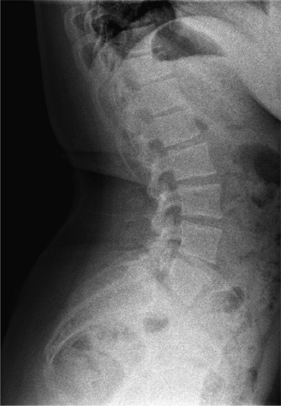
An 18-year-old previously healthy female presented with a six-month history of progressively worsening low back pain radiating to the right lower limb. The pain was initially intermittent but gradually became persistent, resulting in difficulty walking and limitation of daily activities. She denied fever, weight loss, night sweats, cough or previous history of tuberculosis. There was no history of trauma, diabetes mellitus, immunosuppression or other significant comorbidities. Initial evaluation at another institution included a lumbar spine radiograph, which demonstrated Grade I L5 spondylolisthesis (Fig. 1). Her symptoms were attributed to mechanical low back disease, and conservative treatment was initiated. Despite treatment the pain progressively worsened, and she subsequently developed painful swelling over the right gluteal region, prompting further evaluation.
(click to enlarge)
Figure 1. Initial lateral radiograph of the lumbosacral spine demonstrating Grade I anterolisthesis of L5 over S1, which initially supported a mechanical explanation for the patient’s symptoms.
On admission, physical examination demonstrated marked tenderness over the right gluteal region with painful restriction of right hip movement. No neurological deficit was identified. Laboratory investigations showed a normal white blood cell count (6.12 × 10⁹/l) but an elevated erythrocyte sedimentation rate (46 mm/h). C-reactive protein was only mildly elevated (1.3 mg/l); bacterial cultures were negative.
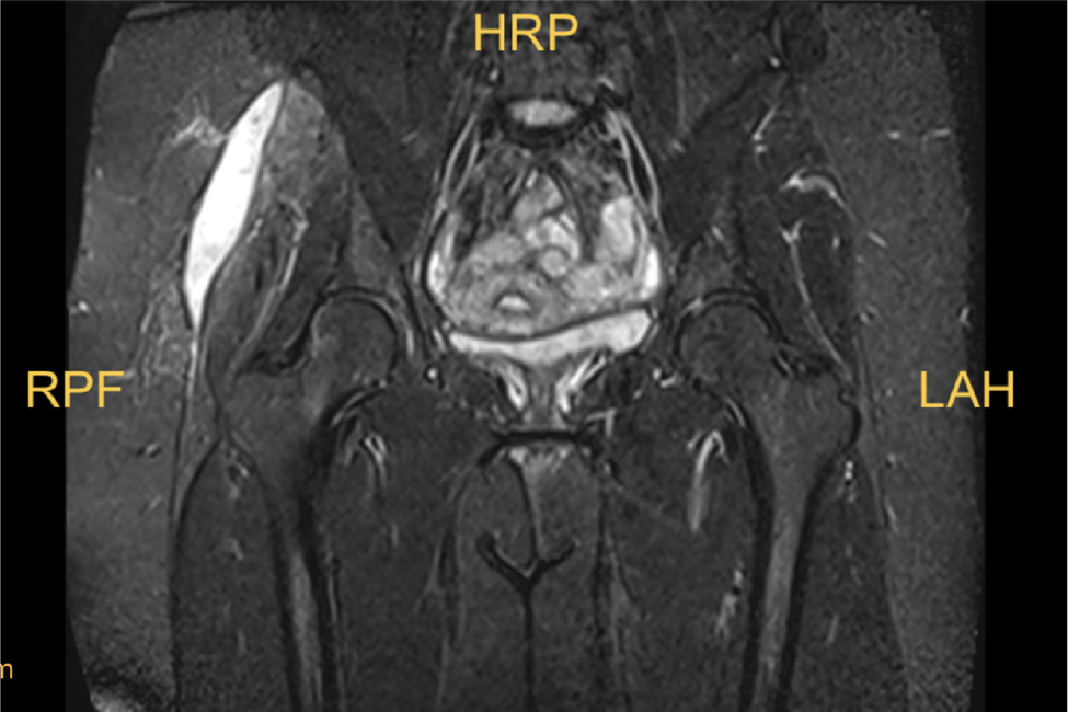
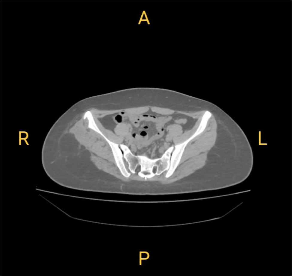
Coronal T2-weighted fat-suppressed magnetic resonance imaging (MRI) of the pelvis demonstrated a well-defined hyperintense intramuscular fluid collection involving the right gluteus medius muscle with surrounding soft-tissue oedema (Fig. 2). Computed tomography (CT) further demonstrated adjacent cortical destruction of the right iliac bone with surrounding soft-tissue extension, raising suspicion of an infective process rather than a purely mechanical disorder (Fig. 3).
(click to enlarge)
Figure 2. Coronal magnetic resonance imaging of the pelvis demonstrating an intramuscular fluid collection with surrounding oedema involving the right gluteus medius muscle, suggestive of an infective process.
(click to enlarge)
Figure 3. Computed tomography of the pelvis demonstrating cortical destruction of the adjacent right iliac bone with surrounding soft-tissue involvement, supporting musculoskeletal infection.
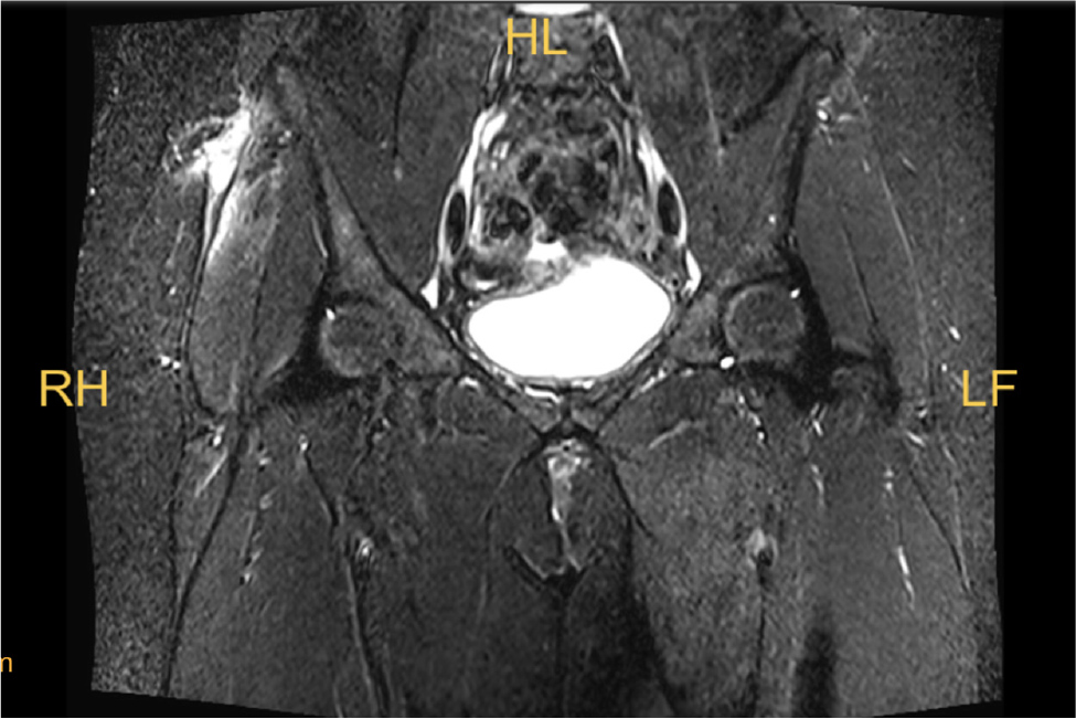
The patient underwent surgical debridement and drainage of the gluteal lesion. However, during follow-up she developed postoperative wound dehiscence, with persistent discharge and sinus tract formation. Repeat laboratory investigations demonstrated a marked inflammatory flare, with C-reactive protein increasing to 85.9 mg/l, while the white blood cell count remained within the normal range (8.93 × 10⁹/l). Repeat MRI demonstrated persistent inflammatory changes with residual collection in the right gluteal region (Fig. 4). Consequently, repeat surgical debridement followed by vacuum sealing drainage was performed to achieve adequate source control.
(click to enlarge)
Figure 4. Follow-up magnetic resonance imaging demonstrating persistent inflammatory changes with residual fluid collection following initial surgical debridement, consistent with postoperative sinus tract formation.
Histopathological examination of the debrided tissue demonstrated necrotic tissue with prominent lymphocytic inflammatory infiltration, consistent with chronic granulomatous infection. Repeated bacterial cultures remained negative. Based on the characteristic imaging findings, histopathological features, intraoperative findings and the clinical picture, a diagnosis of gluteus medius tuberculosis was established, and the patient was commenced on standard anti-tubercular therapy.
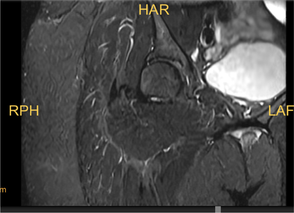
Following surgery and anti-tubercular treatment, the patient’s symptoms improved progressively. Inflammatory markers declined, with the erythrocyte sedimentation rate decreasing to 28 mm/h and C-reactive protein to 7.0 mg/l. Follow-up MRI performed several months later demonstrated near-complete resolution of the gluteal collection and surrounding inflammatory changes without evidence of recurrent disease (Fig. 5). At the most recent follow-up, the patient had resumed normal daily activities without recurrence of pain, wound complications or neurological deficits.
(click to enlarge)
Figure 5. Follow-up magnetic resonance imaging image obtained after completion of treatment demonstrating near-complete resolution of the gluteal lesion and surrounding inflammatory changes without evidence of recurrence.
DISCUSSION
Musculoskeletal tuberculosis is an uncommon manifestation of extrapulmonary tuberculosis, while isolated skeletal muscle involvement remains particularly rare[1]. The relative resistance of skeletal muscle to Mycobacterium tuberculosis, attributed to its abundant blood supply, high lactic acid concentration and paucity of reticuloendothelial tissue, explains the low incidence of primary muscular tuberculosis[1,2]. Consequently, patients often present with non-specific symptoms, and diagnosis is frequently delayed or initially mistaken for pyogenic infection, soft-tissue neoplasm or inflammatory disorders. Recent reviews have also highlighted that constitutional symptoms are absent in many patients, making clinical suspicion particularly challenging[2].
This case demonstrates an additional diagnostic pitfall. The patient had radiographically confirmed Grade I L5 spondylolisthesis, providing a convincing mechanical explanation for chronic low back pain and radicular symptoms. This resulted in diagnostic anchoring; whereby subsequent symptoms were interpreted within the context of an already established spinal diagnosis. Only after persistent pain, progressive gluteal swelling and failure of conservative management was additional cross-sectional imaging performed, ultimately revealing extensive gluteus medius involvement. This sequence illustrates the importance of reassessing the initial diagnosis whenever the clinical course is inconsistent with the expected response to treatment. This case further highlights the importance of avoiding premature diagnostic closure, particularly when the patient’s clinical course is inconsistent with the initial diagnosis despite appropriate treatment.
Published reports describing gluteal tuberculosis remain scarce. Jones and Phillip reported isolated gluteal tuberculosis presenting primarily as gluteal swelling without an associated spinal disorder[3]. Similarly, surgical debridement and anti-tubercular therapy were required to treat a submuscular gluteal abscess arising from sacral tuberculosis[4]. In contrast, the patient had predominant involvement of the gluteus medius muscle together with coexisting lumbar spondylolisthesis, creating a diagnostic dilemma not encountered in these earlier reports. The coexistence of a common degenerative spinal abnormality with an uncommon muscular infection represents the principal educational message of this case rather than the rarity of muscular tuberculosis alone.
MRI proved crucial in establishing the diagnosis by demonstrating intramuscular fluid collection and diffuse inflammatory changes involving the gluteus medius. CT complemented MRI by identifying adjacent cortical destruction of the iliac bone, supporting an infective process rather than isolated mechanical disease. Similar observations have been reported in previous studies, which recognise MRI as the imaging modality of choice for evaluating the extent of musculoskeletal tuberculosis and planning surgical management[1,2]. Histopathological examination in this patient demonstrated necrotic tissue with lymphocytic inflammatory infiltration, but repeated bacterial cultures remained negative. This finding is consistent with the paucibacillary nature of musculoskeletal tuberculosis and emphasises that negative microbiological studies should not exclude the diagnosis when imaging and histopathological findings are strongly suggestive[1].
CONCLUSION
Although anti-tubercular chemotherapy remains the cornerstone of treatment, surgery plays an important role in patients with large abscesses, persistent collections, diagnostic uncertainty or postoperative complications[5]. In this patient, postoperative sinus tract formation required repeat surgical debridement and vacuum sealing drainage before complete clinical recovery was achieved. Subsequent anti-tubercular therapy resulted in progressive clinical improvement, normalisation of inflammatory markers and complete radiological resolution on follow-up MRI. This favourable outcome reinforces previous reports demonstrating excellent outcomes when timely surgical management is combined with appropriate anti-tubercular therapy[4,5].
This case provides an important clinical lesson. In tuberculosis-endemic regions, persistent buttock pain or gluteal swelling should not automatically be attributed to degenerative lumbar spine disease, even when spinal imaging demonstrates a plausible abnormality, such as spondylolisthesis. Failure of symptoms to improve as expected should prompt reconsideration of the diagnosis and early cross-sectional imaging to exclude uncommon conditions, such as muscular tuberculosis.