Acute-on-chronic liver failure revealing autoimmune hepatitis in overlap with inaugural systemic lupus erythematosus
1 Department of Internal Medicine, Hospital Egas Moniz, Unidade Local de Saúde de Lisboa Ocidental, Lisbon, Portugal
2 Department of Internal Medicine, Hospital de São Francisco Xavier, Unidade Local de Saúde de Lisboa Ocidental, Lisbon, Portugal
Corresponding author’s e-mail: dbaptista@ulslo.min-saude.pt
Received: 16/09/2026
Accepted: 12/10/2026
Published: 17/11/2026
Conflicts of Interests: The Authors declare no conflict of interest.
Patient Consent: Written informed consent was obtained from the patient for publication of this report.
This article is licensed under a CC Attribution-NonCommercial-NoDerivatives 4.0 License
How to cite this article: Baptista D, Dâmaso F, Dionísio A, Ferreira P, Duarte J. Acute-on-Chronic Liver Failure Revealing Autoimmune Hepatitis in Overlap with Inaugural Systemic Lupus Erythematosus. EJCRIM 2026;13:doi:10.12890/2026_007531
ABSTRACT
Introduction: Autoimmune hepatitis (AIH) frequently coexists with other autoimmune diseases, including systemic lupus erythematosus (SLE), although it represents a distinct clinical entity rather than a manifestation of these systemic disorders. Given the overlap in autoantibody profiles, the diagnosis of SLE may occasionally be erroneously inferred based solely on serological findings. However, clinically significant hepatic involvement is an uncommon manifestation of SLE.
Case description: The authors present the case of a patient with acute-on-chronic liver failure (ACLF) who was ultimately diagnosed with AIH occurring in overlap with inaugural systemic lupus erythematosus, based on the clinical course, laboratory findings, liver histology and subsequent response to immunosuppressive therapy.
Discussion: The coexistence of multiple plausible aetiologies complicated the initial diagnostic approach, which was subsequently clarified by the development of autoimmune haemolytic anaemia and cytopaenias, allowing the clinical findings to be integrated into a unifying diagnosis.
Conclusion: This case underlines the importance of repeated diagnostic reassessment in patients with acute liver failure and demonstrates that timely recognition of autoimmune disease can improve prognosis and avoid liver transplantation.
KEYWORDS
Systemic lupus erythematosus (SLE), autoimmune hepatitis (AIH), acute-on-chronic liver failure (ACLF), autoimmune haemolytic anaemia, drug-induced liver injury
LEARNING POINTS
- Autoimmune hepatitis (AIH) occurring in overlap with inaugural systemic lupus erythematosus (SLE) may present as acute-on-chronic liver failure (ACLF) and mimic several competing aetiologies.
- Repeated diagnostic reassessment and timely recognition of autoimmune disease can improve prognosis and may avoid liver transplantation.
INTRODUCTION
Acute-on-chronic liver failure (ACLF) is a distinct clinical syndrome characterised by acute hepatic decompensation in patients with underlying chronic liver disease, leading to liver failure, extrahepatic organ dysfunction and high short-term mortality. Early identification of the precipitating event is essential, as timely disease-specific treatment may significantly improve outcomes and prevent liver transplantation. Nevertheless, determining the underlying precipitating cause may be challenging because ACLF encompasses a broad differential diagnosis, frequently requiring systematic exclusion of multiple competing aetiologies[1].
Autoimmune hepatitis (AIH) is a chronic immune-mediated inflammatory liver disease with a heterogeneous clinical presentation, ranging from asymptomatic biochemical abnormalities to acute severe hepatitis and, rarely, acute liver failure. AIH often coexists with other autoimmune diseases, including systemic lupus erythematosus (SLE), although it represents a distinct disease entity rather than a hepatic manifestation of SLE[2].
SLE can be responsible for hepatic abnormalities, however they are usually mild and non-specific; whereas severe hepatitis and acute liver failure are unusual as inaugural manifestations of this disease[3,4]. Given the overlap in autoantibody profiles, the diagnosis of SLE may occasionally be erroneously inferred based solely on serological findings[2].
We present a case of ACLF in which several competing aetiologies were initially considered. However, integration of the subsequent laboratory findings and clinical course ultimately established the diagnosis of inaugural SLE with AIH overlap.
CASE DESCRIPTION
We report the case of a woman in her 50s with obesity, hypertension, recurrent depressive disorder and lumbar disc herniation, whose regular medications included amlodipine/candesartan 5/8 mg daily, lamotrigine 50 mg daily, risperidone 0.5 mg daily, trazodone 300 mg daily and clorazepate as needed. She presented to the emergency department with a two-day history of progressive jaundice. She denied abdominal pain, fever, vomiting, altered bowel habits, pale stools, choluria, recent travel, herbal supplements, wild mushroom ingestion, and consumption of alcohol or other drugs. During the preceding two to three weeks, she had taken paracetamol 1 g three to four doses per day, together with ibuprofen approximately five times daily at unknown dosage. In the days before admission, she had accidentally increased lamotrigine from 50 mg/day to 150 mg/day.
On admission, she was conscious and cooperative with a Glasgow Coma Scale of 14 points. Neurological examination revealed mild psychomotor slowing with occasional episodes of disorientation. She had marked mucocutaneous jaundice and was eupnoeic on room air, with a normal cardiopulmonary examination, a soft and non-tender abdomen, no ascites, no peripheral oedema and no signs of venous thrombosis. Initial laboratory tests showed acute severe liver dysfunction with an international normalised ratio of 2.7 (reference range <1.3), fibrinogen 1.16 g/l (reference range 2-4), albumin 2.7 g/dl (reference range 3.5-5.0), factor V 46% (reference range 70-150), ammonia 76 µmol/l (reference range 11-35), total bilirubin 14.8 mg/dl (reference range <1.40), conjugated bilirubin 11.3 mg/dl (reference range <0.3), aspartate aminotransferase (AST) 686 IU/l (reference range <40), alanine aminotransferase (ALT) 261 IU/l (reference range <41), gamma-glutamyl transferase 120 IU/l (reference range 10-71), alkaline phosphatase 68 IU/l (reference range 40-130), blood urea nitrogen (BUN) 15 mg/dl (reference range 17-49); creatinine 1.09 mg/dl (reference range 0.7-1.2) and lactate dehydrogenase (LDH) 308 IU/l (reference range 135-225). Paracetamol was 8 μg/ml and viral studies including serologies for hepatitis A, B and C, cytomegalovirus and human immunodeficiency virus testing were negative. Abdominal ultrasound showed irregular hepatic contours, coarse echotexture, unobstructed gallstones and mild splenomegaly, suggesting underlying chronic liver disease. Supporting this, an abdominal ultrasound performed a few years earlier had documented hepatic steatosis.
The patient fulfilled criteria for Grade 2 ACLF and was admitted to the intermediate medical care unit on the same day. Given the recent history of repeated supratherapeutic paracetamol exposure, frequent non-steroidal anti-inflammatory drug consumption and accidental lamotrigine dose escalation, the initial assumed diagnosis was drug-induced liver injury—particularly subacute paracetamol toxicity—and N-acetylcysteine therapy was immediately initiated.
Despite the initial suspicion of drug-induced liver injury, the diagnostic work-up rapidly revealed features suggestive of systemic autoimmune disease. Viral hepatitis A, B, C and E, HIV, syphilis, Wilson disease, alpha-1 antitrypsin deficiency and vascular causes of liver disease were excluded. Although ferritin and transferrin saturation were elevated, the overall context favoured inflammatory liver injury rather than primary haemochromatosis. Autoimmune testing demonstrated marked polyclonal hypergammaglobulinaemia with predominance of IgG (5,150 mg/dl; reference range 600-1,500), antinuclear antibody titres >1:1,280, anti-double-stranded DNA antibodies of 916.1 IU/ml (reference range <30), hypocomplementaemia with C3 48.8 mg/dl (RR 90–180) and C4 5.4 mg/dl (reference range 10-40), positive antimitochondrial antibodies, negative anti-LKM, negative anti-SLA/LP, strongly positive anti-Sp100 antibodies, weakly positive anti-PML antibodies, negative anti-histone antibodies and inconclusive smooth muscle antibodies.
The marked hypergammaglobulinaemia and high-titre antinuclear antibody positivity raised the possibility of AIH. Consequently, methylprednisolone 80 mg/day (1 mg/kg) was initiated seven days after admission. In parallel, due to the coexistence of positive antimitochondrial and anti-sp100 antibodies, primary biliary cholangitis (PBC) overlap was considered and ursodeoxycholic acid 1,250 mg/day was therefore introduced on the same day and subsequently maintained. Corticotherapy was tapered to prednisone 60 mg/day the following week (12 days after admission).
However, the subsequent evolution created considerable diagnostic uncertainty. Although aminotransferase levels progressively decreased, liver synthetic function failed to improve, as shown in Table 1, and urgent liver transplantation was becoming a realistic possibility. The hepatology team recommended discontinuation of corticosteroids on day 16 of hospitalisation due to concern that ongoing corticosteroid exposure might increase surgical complications should liver transplantation become necessary.
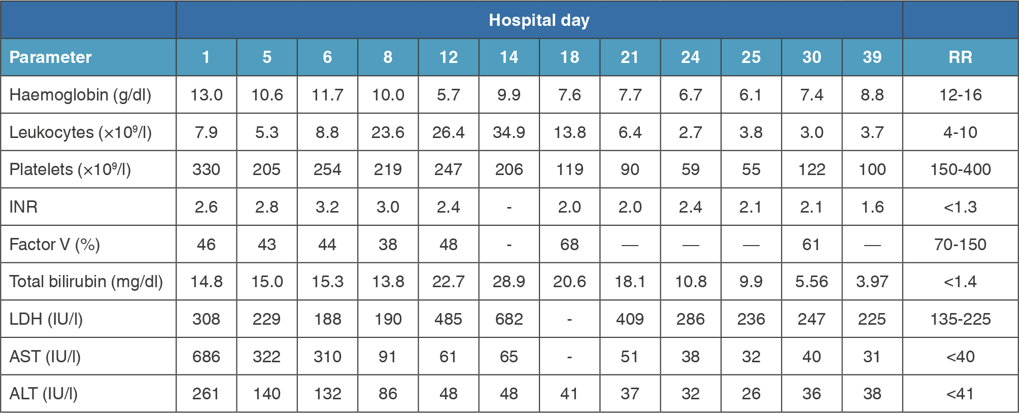
(click to enlarge)
Table 1. Evolution of haematological and liver biochemistry parameters during hospitalisation.
Abbreviations: INR, international normalised ratio; ALT, alanine transaminase; AST, aspartate aminotransferase; LDH, lactate dehydrogenase; RR, reference range.
A decisive turning point occurred by day 12 after admission. The patient developed severe anaemia, with haemoglobin decreasing from 13.0 g/dl on admission to 5.7 g/dl. Simultaneously, total bilirubin increased to 22.7 mg/dl despite AST decreasing to 61 IU/l and ALT to 48 IU/l. This dissociation between improving hepatocellular injury and worsening jaundice suggested an additional mechanism contributing to hyperbilirubinaemia. Further investigation demonstrated reticulocytosis of 11.1% (reference range 0.5-2.5), haptoglobin <10 mg/dl, elevated LDH, positive direct antiglobulin test for IgG and presence of cold agglutinins, establishing the diagnosis of mixed warm and cold autoimmune haemolytic anaemia. One unit of warmed red blood cells was transfused due to severe and symptomatic anaemia.
The emergence of autoimmune haemolytic anaemia, together with evolving thrombocytopaenia, leukopenia, hypocomplementaemia, markedly elevated anti-dsDNA antibodies and hypergammaglobulinaemia, fundamentally changed the diagnostic reasoning. Liver disease could no longer be interpreted as an isolated process and appeared to represent part of a systemic autoimmune disorder, supporting the diagnosis of inaugural SLE with hepatic and haematological involvement. Neuropsychiatric involvement was excluded by brain magnetic resonance imaging (MRI). Antiphospholipid testing was negative, including lupus anticoagulant, anticardiolipin IgG/IgM and anti-beta-2 glycoprotein IgG/IgM. Mild proteinuria was present, but 24-hour urinary protein was below 500 mg/day without significant albuminuria, excluding relevant lupus nephritis at that stage.
A transjugular liver biopsy was performed, and the result became available on the 24th day of hospitalisation. Histology showed global architectural distortion due to confluent hepatocellular necrosis, mixed portal and lobular inflammatory infiltrates with rare plasma cells and eosinophils, activated macrophages, focal ballooning, canalicular and ductular cholestasis, ductular proliferation and intermediate phenotype hepatocytes. There was no steatosis or granuloma, and histochemical stains for copper and alpha-1 antitrypsin were negative. Fibrosis could not be reliably assessed because of extensive necrosis. The final interpretation was moderate-to-severe acute hepatitis with a differential diagnosis including AIH, drug-induced injury and viral hepatitis. However, the pathological pattern was considered not typical of paracetamol toxicity and viral hepatitis had been excluded. AIH, probably with areas of chronic involvement, became the leading diagnosis.
Following integration of the histological findings with the evolving autoimmune and haematological phenotype, and after multidisciplinary discussion with the liver transplant unit, the diagnosis of SLE with AIH overlap was considered the unifying diagnosis for the patient’s presentation.
Consequently, disease-directed immunosuppressive therapy was reintroduced on that day, with high-dose methylprednisolone (80 mg/day) initially, followed by prednisolone 80 mg/day, later tapered to 60 mg/day and subsequently to 40 mg/day. Hydroxychloroquine 400 mg/day was also initiated concomitantly with mycophenolate mofetil 1,000 mg twice daily, which was later escalated to 1,500 mg twice daily.
Following the introduction of targeted immunosuppressive therapy, liver synthetic function gradually recovered with progressive reduction in the international normalised ratio and a sustained decline in total bilirubin levels. Haematological recovery was also observed, with gradual improvement in haemoglobin concentration, leukocyte count and platelet count. Evidence of active haemolysis progressively resolved, with stabilisation of haemoglobin levels and normalisation of lactate dehydrogenase. Clinically, jaundice regressed, hepatic encephalopathy resolved and functional status progressively improved with physiotherapy. Ultimately, liver transplantation was no longer considered necessary. On 9 June, repeat assessment showed a decrease in anti-double-stranded DNA antibody levels to 168.5 IU/ml; however, hypocomplementaemia persisted (C3 49 mg/dl and C4 6.9 mg/dl). The patient was discharged on 17 June in a stable clinical condition. Follow-up outpatient evaluation over the subsequent weeks demonstrated progressive clinical and biochemical improvement.
DISCUSSION
This case illustrates the diagnostic difficulty of ACLF when multiple plausible aetiologies coexist[1]. At presentation, drug-induced liver injury was the most immediate and appropriate working hypothesis. However, several findings argued against paracetamol toxicity as the sole explanation. First, the paracetamol level was low, and the clinical presentation suggested subacute rather than classic acute massive ingestion. Second, imaging demonstrated irregular liver contours and coarse echotexture, indicating pre-existing chronic liver disease. Third, aminotransferase levels declined rapidly while hyperbilirubinaemia and hepatic synthetic dysfunction persisted, an evolution not fully explained by resolving paracetamol toxicity. Fourth, liver biopsy was not typical of paracetamol-induced liver injury and instead raised AIH as the leading diagnostic possibility. Finally, the emergence of autoimmune haemolytic anaemia and evolving cytopaenias could not be explained by paracetamol toxicity[5].
Drug-induced autoimmune-like hepatitis also had to be considered, given that lamotrigine is a recognised cause of idiosyncratic liver injury—often in the context of hypersensitivity syndromes, and the patient had accidentally increased her dose[6]. Nevertheless, this diagnosis was considered unlikely because lamotrigine had been used chronically, the dose escalation was brief, and the subsequent clinical course was not consistent with this hypothesis.
PBC overlap was suspected because of positive antimitochondrial and anti-Sp100 antibodies, justifying the early introduction of ursodeoxycholic acid. However, several findings argued against PBC as the primary diagnosis. The biochemical profile was predominantly hepatocellular rather than cholestatic, alkaline phosphatase remained within the normal range and liver biopsy did not demonstrate histological features supporting PBC[7].
The severity of hepatic synthetic dysfunction, together with markedly elevated IgG levels and high-titre antinuclear antibody positivity, strongly supported the diagnosis of AIH[2]. An initial biochemical response to methylprednisolone was observed, with progressive improvement in aminotransferase levels. However, following tapering to prednisone 60 mg/day, the patient developed autoimmune haemolytic anaemia and pancytopaenia, suggesting an underlying systemic autoimmune disorder. This clinical and biochemical evolution indicated that the patient’s presentation was best explained by inaugural SLE with AIH overlap rather than two unrelated disease processes[2-4]. The subsequent improvement in liver synthetic function, bilirubin, haematological parameters and anti-dsDNA titres following targeted immunosuppressive treatment further strengthened this interpretation[2,3].
An additional consideration is that the patient may also have had underlying metabolic dysfunction-associated steatotic liver disease. She had obesity and a previous abdominal ultrasound demonstrating hepatic steatosis several years before presentation, supporting pre-existing metabolic liver disease. Current EASL guidelines recognise that AIH may coexist with metabolic dysfunction-associated steatotic liver disease and that this overlap is increasingly encountered in clinical practice. Management should additionally include lifestyle intervention, weight reduction and optimisation of metabolic risk factors[2].
CONCLUSION
This case highlights that AIH in overlap with inaugural SLE may present as ACLF, creating a challenging differential diagnosis and requiring repeated diagnostic reassessment. The development of autoimmune haemolytic anaemia and evolving cytopenias was pivotal in recognising the underlying systemic autoimmune disease. Integration of the clinical, serological and histological findings supported the final diagnosis and enabled timely initiation of targeted immunosuppressive therapy. Early referral to a liver transplant centre remains essential in severe autoimmune liver disease; however, prompt recognition and treatment of the underlying immune-mediated process may lead to clinical recovery and ultimately avoid liver transplantation[1,2].