DOI: 10.12890/2025_005391
Transverse myelitis successfully treated with posterior decompression followed by secretome and mesenchymal stem cell therapy
Authors
 ahmad.faried@unpad.ac.id
Keywords
Transverse myelitis (TM), longitudinally extensive transverse myelitis (LETM), secretome and mesenchymal stem cells therapyAbstract
Introduction: Transverse myelitis (TM) is a rare spinal cord disorder caused by inflammation. Usually, this occurs as a complication of infection or autoimmune disease; however, idiopathic causes such as vaccinations have been reported. There have been no studies that include the use of stem cells for TM in an inpatient setting. We present a case demonstrating the efficacy of stem cell therapy in the treatment of TM.
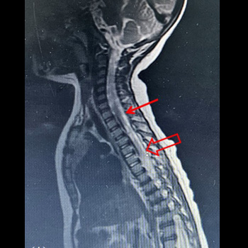
Case presentation: A 5-month-old boy developed TM (he abruptly developed paraplegia, paraesthesia of both legs, urinary retention) 7 days after he received the pneumococcal conjugate vaccine (PCV). Laboratory tests were all negative except for evidence of recent PCV vaccination. A paediatric neurologist confirmed the diagnosis when the boy was 2.5 years old. Magnetic resonance imaging of thoracic spine showed hyperintense central and dorsal cord abnormalities beginning at the T2 level and extending into the thoracic cord at T6. The pattern was consistent with TM. Decompression laminectomy was performed. The patient was then given mesenchymal stem cell (MSC)-derived secretome and umbilical cord mesenchymal stem cell (UCMSC) administered intrathecally (3× within interval 4 weeks). After the first, treatment he began to show significant improvement in terms of motor function and the patient also showed increasing in autonomic function especially in erectile function. After the 2nd treatment there was adequate muscle response of the lower limbs, and his medical rehabilitation specialist trained him to do standing exercises. After the 3rd treatment there was adequate muscle response of the lower limbs, and his medical rehabilitation specialist trained him to walk.
To the best of our knowledge this is the first reported case of TM treated with stem cells in Indonesia. The pathophysiology of TM is an inflammatory disorder of the spinal cord that damages the myelin sheath of nerve fibres. Two primary therapies for TM are glucocorticoids and plasma exchange. Further, treatment options such as with secretome and UCMSCs promote both microenvironment correction and myelin regeneration.
To the best of our knowledge our case is the first reported case of TM in Indonesia treated with stem cells.
Conclusion: Our case demonstrates the potential of stem cell therapy as an alternative treatment modality in a case there was no other optional therapy for longitudinal extensive TM.
References
- Kitley JL, Leite MI, George JS, Palace JA. The differential diagnosis of longitudinally extensive transverse myelitis. Mult Scler 2012;18:271-285. doi: 10.1177/1352458511406165
- Simone CG, Emmady PD. Transverse Myelitis. [Updated 2022 Nov 15]. In: StatPearls. Treasure Island (FL): StatPearls Publishing; 2025 January. Link Available from: https://www.ncbi.nlm.nih.gov/books/NBK559302/
- Transverse Myelitis Consortium Working Group. Proposed diagnostic criteria and nosology of acute transverse myelitis. Neurology 2002;59:499-505. doi: 10.1212/wnl.59.4.499
- Cortese I, Chaudhry V, So YT, Cantor F, Cornblath DR, Rae-Grant A. Evidence-based guideline update: Plasmapheresis in neurologic disorders: report of the Therapeutics and Technology Assessment Subcommittee of the American Academy of Neurology. Neurology 2011;76:294-300. doi: 10.1212/WNL.0b013e318207b1f6
- Peng C, Li Y, Lu L, Zhu J, Li H, Hu J. Efficient One-Step Induction of Human Umbilical Cord-Derived Mesenchymal Stem Cells (UC-MSCs) Produces MSC-Derived Neuro-spheres (MSC-NS) with Unique Transcriptional Profile and Enhanced Neurogenic and Angiogenic Secretomes. Stem Cells Int 2019;2019:9208173. doi: 10.1155/2019/9208173
- Fisher-Shoval Y, Barhum Y, Sadan O, Yust-Katz S, Ben-Zur T, Lev N, et al. Transplantation of placenta-derived mesenchymal stem cells in the EAE mouse model of MS. J Mol Neurosci 2012;48:176-184. doi: 10.1007/s12031-012-9805-6
- Zhang J, Li Y, Lu M, Cui Y, Chen J, Noffsinger L, Elias SB, Chopp M. Bone marrow stromal cells reduce axonal loss in experimental autoimmune encephalomyelitis mice. J Neurosci Res 2006;84:587-595. doi: 10.1002/jnr.20962
- Lotfy A, Ali NS, Abdelgawad M, Salama M. Mesenchymal stem cells as a treatment for multiple sclerosis: A focus on experimental animal studies. Rev Neurosci 2020;31:161-179. doi: 10.1515/revneuro-2019-0040
- Medina R, Derias AM, Lakdawala M, Speakman S, Lucke-Wold B. Overview of emerging therapies for demyelinating diseases. World J Clin Cases 2024;12:6361-6373. doi: 10.12998/wjcc.v12.i30.6361
- Vaquero J, Zurita M, Rico MA, Aguayo C, Bonilla C, Marin E, et al. Intrathecal administration of autologous mesenchymal stromal cells for spinal cord injury: safety and efficacy of the 100/3 guideline. Cytotherapy 2018;20:806-819. doi: 10.1016/j.jcyt.2018.03.032
- Yang Y, Pang M, Du C, Liu ZY, Chen ZH, Wang NX, et al. Repeated subarachnoid administrations of allogeneic human umbilical cord mesenchymal stem cells for spinal cord injury: a phase 1/2 pilot study. Cytotherapy 2021;23:57-64. doi: 10.1016/j.jcyt.2020.09.012